
Prediction of ACL Strain Using Combined CT and Biplane Fluoroscopy: A Sensitivity Analysis
Currently, there is no practical way to directly measure anterior cruciate ligament (ACL) strain in vivo during dynamic activities. Placing a transducer directly on the ACL is inherently invasive and mathematical models require substantial development and are difficult to apply to a specific subject. Precise measurement of bony motion using biplane fluoroscopy, developed at the Steadman Hawkins Research Foundation under the direction of Drs. Richard Steadman and Mike Torry, may allow the prediction of in vivo ligament strains based on the location of their bony origin and insertions.
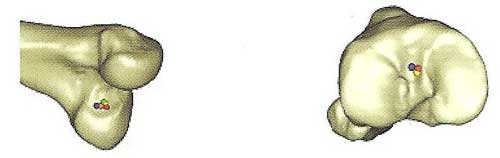
Recently, Drs. Torry, Erik Giphart and Kevin Shelburne, used the biplane fluoroscopy system to measure the bony motion of six knees from six male subjects as they performed a drop landing from a height of 40 cm. For each knee, the origin and insertion of the ACL was selected based on detailed anatomic descriptions (Fig. 1). ACL length was measured as the straight line distance between the center of the femoral origin and insertion sites of the ligament. A sensitivity analysis was performed to determine the practicality of predicting ACL length and strain from bony motion measured with biplane fluoroscopy.
During landing from a height of 40 cm, the length of the ACL decreased from toe-touch to maximum knee flexion. Baseline peak ACL strain was dissimilar between the subjects. Small changes in the selected origin and insertion site of the ligament had little effect on the length predictions for each subject. In summary, use of this method to show inter-subject differences between activities, for instance, is supported by these results. However, the calculation of strain from these measurements was overly sensitive to small changes in the selected origin and insertion sites. For half the subjects, the 1.5mm variations produced changes in strain spanning the normal function of the ligament. Also, these methods may be sufficient for inter-subject comparison of predicted strain; however, prediction of absolute strain may require more precise modeling of the ligament origin and insertion sites and substance of the ACL.